Mammography
Mammography is a fundamental examination for assessing the health of the breast. It uses ionizing radiation to deliver a morphological image of the breast that can detect breast lesions or areas of structural distortion. Among the breast lesions recognized by mammography there are those of tumorous origin that present themselves in the form of nodular opacities with irregular margins and polymorphic micro-calcifications.
Thanks to technological evolution, the classic analogue mammography has evolved into Tomosynthesis mammography. The latter acquires multilayer images of the organ under examination in order to increase its resolution and consequently its diagnostic capacity. Tomosynthesis uses an extremely low and harmless radiogenic dose but has the advantage of being very sensitive to the recognition of very small tumorous lesions and specific for the exclusion of false positives.
Mammography is an examination recommended for all women who have reached 40 years of age, the age of highest incidence of breast cancer. In women with particularly dense breasts, a check-up every 12 months and/or an ultrasound examination that compensates for the diagnostic evaluation is recommended. In women with less dense breasts or who do not have any indication for breast cancer, an interval between the two tests is recommended that can also be extended to two years.
To date, mammography is the main test of choice for the prevention of breast cancer. The early diagnosis of this tumour is fundamental for its positive resolution. However, when the detection occurs at an advanced stage, the possibility of treatment is more limited. For this reason, all women from the age of 40 to over 70 years should undergo mammography.
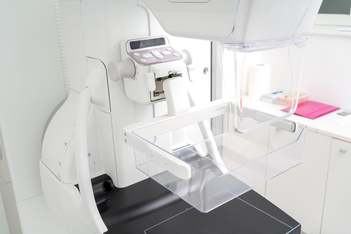
This examination does not require anaesthesia, lasts only a few minutes and is not particularly painful. The instrument used - the mammograph - projects an X-ray beam directly onto the breast, which is positioned on a detector (support surface); the breast is compressed by means of a plastic plate in order to immobilize it.
The compression of the breast allows 1) the acquisition of a high quality X-ray images; 2) the reduction of the thickness of the breast to which follows the use of lower radiation. At each standard mammographic examination, to ensure an accurate view of the organ, two projections per breast are acquired: one cranio-caudal and one oblique mid-lateral.
Finally, mammographic examinations performed over time can be compared in order to observe variations. Stability over time is another index of benignity and allows the exclusion of any suspicion.